

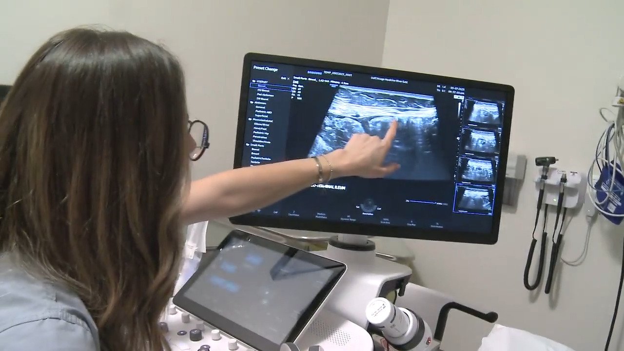
The Clinical Shift Toward Point-of-Care Bowel Imaging
Gastroenterology practices increasingly rely on intestinal ultrasound as a front-line diagnostic tool because it bypasses the logistical bottlenecks of traditional radiology departments. Inflammatory bowel disease encompasses chronic, relapsing conditions like Crohn’s disease and ulcerative colitis that demand frequent physiological reassessments to maintain clinical remission. Traditional cross-sectional imaging methods often involve long scheduling delays, complex patient preparation protocols, and cumulative ionizing radiation, making them less than ideal for longitudinal disease tracking over a patient’s lifetime. Intestinal ultrasound transforms this paradigm by bringing real-time diagnostic capability directly into the outpatient clinic setting, allowing physicians to visualize tissue changes immediately during a routine consultation without administrative friction.
Furthermore, decentralized medical care models continue to evolve across various healthcare sectors, much like examining broader care frameworks such as exploring home-based elder support alternatives, which highlights how bringing specialized diagnostic and supportive services closer to the patient substantially improves overall comfort and reduces travel burdens for vulnerable populations. By integrating point-of-care sonography directly into routine clinic visits, gastroenterologists empower patients with immediate clinical answers and foster collaborative treatment planning that adapts seamlessly to daily life.
Eliminating Radiation Risks During Longitudinal Care
Patients managing lifelong gastrointestinal disorders must undergo regular diagnostic imaging to evaluate therapeutic efficacy, identify subclinical complications, and monitor disease progression over many decades. Computed tomography scans provide excellent anatomical detail of the entire abdominal cavity but expose vulnerable tissues to substantial doses of ionizing radiation, creating cumulative safety concerns over long-term management schedules. Magnetic resonance imaging successfully eliminates radiation exposure, yet high operational costs, restricted machine availability, and lengthy scan times present significant logistical barriers for routine follow-ups in busy clinical environments. Intestinal ultrasound resolves these dilemmas completely by utilizing harmless high-frequency sound waves, enabling clinicians to perform repeat examinations as often as clinically necessary without any biological hazard or cumulative tissue risk.
Quantifying Bowel Wall Thickness and Vascular Signals
Clinical assessment via intestinal ultrasound focuses on precise physical parameters, including bowel wall thickness, wall layer stratification, and mesenteric blood flow velocity. A healthy intestinal wall typically measures under two millimeters across from the lumen to the serosa, whereas active inflammatory flare-ups cause the muscularis propria and submucosa to swell significantly beyond normal diagnostic thresholds. Gastroenterologists routinely measure wall thickness exceeding three millimeters to confirm active disease segments and localize inflammatory activity with high spatial resolution. Furthermore, power Doppler signal analysis detects hyperemic blood flow within the affected tissue layers, providing a direct physiological marker of acute inflammation before clinical symptoms fully manifest or laboratory inflammatory markers spike.
Evaluating Transmural Healing and Biological Response
Modern inflammatory bowel disease management targets deep transmural healing rather than relying solely on the temporary resolution of external clinical symptoms like abdominal cramping or stool frequency. Achieving transmural healing means that inflammation resolves across all distinct layers of the intestinal wall, rather than just on the mucosal surface visible during standard endoscopic procedures. When physicians introduce advanced biologic therapies, small molecule inhibitors, or targeted immunomodulators, serial intestinal ultrasound scans map out whether bowel wall thickness and vascular signals are steadily returning to normal baseline levels. This objective, non-invasive feedback loop confirms whether a specific pharmaceutical intervention is working effectively deep within the gastrointestinal tract, guiding confident clinical decisions regarding drug continuation or dose optimization.
Patient Comfort, Minimal Preparation, and Accessibility
Patient compliance improves dramatically when diagnostic procedures cause no physical pain and require minimal preparation beforehand compared to traditional invasive alternatives. Unlike diagnostic colonoscopies that necessitate aggressive bowel cleansing agents, strict dietary restrictions, and professional sedation, a standard intestinal ultrasound scan requires little more than comfortable clothing and occasional short-term fasting to reduce interfering bowel gas. Individuals lie relaxed on an examination table while a trained sonographer or gastroenterologist applies warm conductive gel and glides a handheld transducer gently across the anterior and lateral abdominal wall. This gentle, interactive examination format reduces procedural anxiety and encourages patients to attend scheduled surveillance appointments faithfully, ensuring that recurring inflammation or early relapse is caught before severe complications develop.
Common Practical Inquiries About Bowel Sonography
Does intestinal ultrasound require any special preparation before the appointment?
Most standard intestinal ultrasound examinations require little to no special preparation, though clinicians occasionally ask patients to fast for a few hours prior to the scheduled scan to reduce bowel gas interference and enhance image clarity.
How long does a typical intestinal ultrasound procedure take?
A typical intestinal ultrasound scan takes between fifteen and thirty minutes to complete, depending on the anatomical extent of the bowel segments that require examination and the complexity of the inflammation.
Can intestinal ultrasound completely replace colonoscopy for inflammatory bowel disease?
While intestinal ultrasound is exceptionally useful for tracking bowel wall thickness and transmural healing without radiation, colonoscopy remains essential for direct mucosal biopsy, dysplasia surveillance, and detailed surface visualization.
Is intestinal ultrasound painful for patients experiencing an active flare-up?
The procedure is generally painless and well-tolerated, though slight, temporary discomfort may occur if the transducer applies mild pressure directly over an area of acute transmural inflammation.
How often should patients with inflammatory bowel disease undergo ultrasound monitoring?
The exact frequency of ultrasound monitoring is determined by your gastroenterologist based on your specific treatment plan, current disease stability, symptom history, and response to biologic medications.
Does insurance typically cover intestinal ultrasound examinations?
Coverage varies significantly by insurance provider and geographic region, so patients should always verify specific diagnostic benefits and prior authorization requirements directly with their carrier prior to the appointment.
Disclaimer: The medical information provided in this article is for educational purposes only and should not replace professional medical advice, clinical diagnosis, or specialized treatment. Always consult your gastroenterologist or qualified healthcare provider regarding any questions about your condition or ongoing treatment plan.

Passionate content writer and savvy blog publisher, Aamir crafts compelling stories and insightful articles that captivate and inform. With a knack for blending creativity and strategy, they bring fresh perspectives to every piece. Dive into their world of words and discover content that resonates.


